In microbiology, a simple stain lets you quickly see the basic shape and size of microorganisms by using just one dye, like methylene blue or crystal violet. You’ll apply the stain to a prepared smear, which highlights cells against the background for easy viewing under a microscope. This technique is fast, easy, and great for beginners, though it won’t show internal details or differentiate types. Keep going to uncover more about how it works and where it’s used.
Key Takeaways
- A simple stain uses a single dye to color microorganisms for easier visibility under a microscope.
- It highlights basic bacterial shapes like rods, spheres, and spirals without showing internal details.
- Common dyes include methylene blue, crystal violet, and safranin, which bind to negatively charged cell surfaces.
- The process involves applying dye to a heat-fixed smear, rinsing, and then observing the stained cells.
- Simple staining is quick, cost-effective, and ideal for preliminary morphology assessment but cannot differentiate bacterial types.
Definition and Purpose of Simple Stains
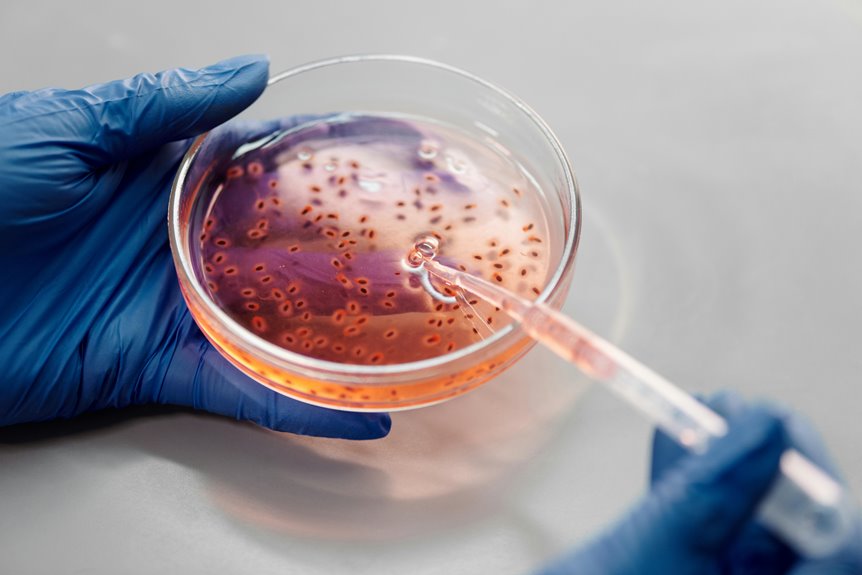
A simple stain uses a single dye to color microorganisms, making them easier for you to see under a microscope. When you apply this stain, it highlights the cells by increasing contrast between the microorganisms and their background.
This method helps you identify basic shapes like rods, spheres, or spirals, and observe cell size and arrangement. You won’t get details about internal structures with a simple stain, but it’s perfect for quickly evaluating morphology.
Since it’s straightforward and fast, you can use it as a first step before more complex staining techniques. By mastering simple stains, you’ll build a solid foundation for understanding microbial form and preparing samples for further analysis.
Simple staining makes microscopic observation clearer and more effective for your studies.
Common Dyes Used in Simple Staining
To make microorganisms stand out clearly under the microscope, you’ll need the right dyes for simple staining. Commonly, basic dyes like methylene blue, crystal violet, and safranin are used because they carry a positive charge.
This positive charge allows them to bind easily to the negatively charged surfaces of bacterial cells, highlighting their shape and arrangement. Methylene blue is popular for its ease of use and clear contrast. Crystal violet offers a deep purple color, making details more visible.
Positively charged dyes like methylene blue and crystal violet highlight bacterial shapes with clear, vivid contrast.
Safranin, a red dye, is often chosen when you want a bright, contrasting color. These dyes are straightforward, reliable, and provide quick results, helping you observe microorganisms with clarity and simplicity.
Choosing the right dye depends on the specific microorganisms and the details you want to emphasize.
Step-by-Step Procedure for Simple Staining
Although simple staining may seem straightforward, following a precise step-by-step procedure guarantees you get clear, detailed images of microorganisms every time.
First, prepare a clean glass slide and place a small drop of water on it if you’re using a solid culture. Next, transfer a tiny amount of your bacterial sample to the slide and spread it thinly. Allow the smear to air dry completely.
Then, heat-fix the sample by quickly passing the slide through a flame to kill and adhere the cells. After that, cover the smear with your chosen simple stain, such as methylene blue, and let it sit for about one minute.
Rinse the slide gently with distilled water to remove excess dye, then blot dry with bibulous paper. Finally, examine the stained slide under a microscope.
Advantages of Using Simple Stains
Why choose simple stains when you want to quickly observe microorganisms? Simple stains offer a fast and effective way to see cell shapes, sizes, and arrangements without complicated steps.
You don’t need special reagents or extensive preparation, so you can get results in minutes. These stains increase contrast by coloring the cells, making it easier for you to examine them under a microscope.
Since the process is straightforward, it’s perfect for beginners or when you’re screening multiple samples. Simple stains also help you identify basic morphological features that are essential for classifying bacteria.
Plus, they require minimal equipment and materials, keeping costs low. Overall, simple staining is a reliable, efficient method to gain clear insights into microbial structure quickly.
Limitations of Simple Staining Techniques
While simple stains help you see basic cell features quickly, they don’t provide detailed information about a microorganism’s internal structures or differentiate between types of cells.
You won’t be able to distinguish between Gram-positive and Gram-negative bacteria or identify specific cellular components like spores or capsules using simple staining. Additionally, simple stains often result in a uniform color, which can make it hard to observe cell arrangements or subtle morphological differences.
If your sample contains mixed populations, simple staining won’t help you separate them. Also, because simple stains rely on basic dyes, they may not bind well to all cell types, potentially causing inconsistent results.
Understanding these limitations helps you choose the right staining technique for your microbiological analysis.
Differences Between Simple and Differential Stains
Because simple and differential stains serve different purposes, understanding their distinctions is essential for effective microscopy.
When you use a simple stain, you apply one dye to color all cells uniformly, which helps you observe cell shape, size, and arrangement quickly. In contrast, differential stains use multiple dyes to highlight differences between cell types or structures, allowing you to distinguish between bacteria like Gram-positive and Gram-negative.
Simple stains are straightforward and fast, but they don’t provide detailed information about cell composition.
Simple stains offer quick visualization but lack detailed insights into cellular composition.
Differential stains, while more complex, give you deeper insights by revealing structural or chemical variations. Knowing when to choose a simple stain versus a differential one helps you get the right information from your microscopic analysis.
Applications of Simple Stains in Microbiology
Simple stains play a key role in microbiology by providing a quick and effective way to observe basic cell characteristics. When you apply a simple stain, you instantly enhance the visibility of microorganisms, making it easier to study their shape, size, and arrangement.
You’ll find simple stains especially useful in these situations:
- Identifying the general morphology of bacteria
- Counting cells in a sample
- Observing bacterial arrangements like clusters or chains
- Differentiating between live and dead cells with specific dyes
- Preparing samples for further microscopic analysis
Tips for Achieving Optimal Staining Results
To get the best results from your staining process, you need to follow a few key steps carefully.
First, make sure your bacterial smear is thin and evenly spread; thick smears can lead to uneven staining and poor visualization. Always air-dry the smear completely before heat-fixing to prevent distortion.
Use the stain at the recommended concentration and apply it for the right amount of time—usually around one minute—to avoid over- or under-staining. Rinse gently with water to remove excess dye without washing away the sample.
Finally, let your slide air dry before observing under the microscope. By paying close attention to these details, you’ll enhance contrast and clarity, making it easier to identify cell morphology accurately.
Frequently Asked Questions
How Long Should a Simple Stain Slide Be Stored Before Examination?
You should examine a simple stain slide as soon as possible after preparation to get the best results. If you must store it, keep it dry and examine within 24 hours to avoid fading or distortion.
Can Simple Stains Be Used on Live Bacterial Cultures?
You can’t use simple stains on live bacterial cultures because the staining process usually kills the cells. Simple stains require fixed, dead cells to properly absorb the dye and reveal their shape and arrangement under the microscope.
What Safety Precautions Are Necessary When Handling Staining Dyes?
Think of staining dyes as magical potions—handle them with gloves and goggles, avoid inhaling fumes, and work in a ventilated space. You wouldn’t want those potions to harm you while revealing hidden secrets, right?
How Does Temperature Affect the Staining Process?
You’ll find that temperature influences stain uptake; higher temperatures speed up dye penetration but can distort cell structures. Cooler temps slow staining, so maintaining an ideal temperature guarantees accurate, consistent results without damaging your samples.
Are Simple Stains Effective for All Types of Microorganisms?
Simple stains work well for most microorganisms because they highlight cell shape and size. However, they don’t differentiate between species or cell structures, so for detailed analysis, you’ll need more specific staining techniques.